
It is a medical test that doctors use to diagnose the disease.
CT takes cross-sectional pictures of the inside of the body using special x-rays and a computer. More detailed clear images are obtained compared to normal x-ray examination.
CT is used for almost all parts of the body. The main ones are lung, abdomen, brain, spine, vessels, heart and intestines.
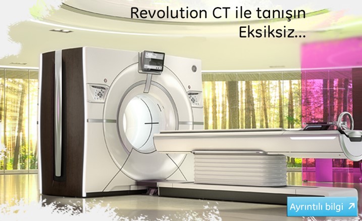
Meet Revolution * CT
The ability to examine the inside of the human body noninvasively is a it is a modern day miracle. This is for CT imaging professionals like you and made possible for millions of people with the work of their doctors. a miracle coming.
As with any technology that evolves over time, sometimes just taking the next step is not enough. To a point like that We get where we want with just one big leap we can reach.
Every patient's needs are different.
Your CT system meets the most demanding needs of all your patients. How would it be if he could afford it? Well, clinic in all departments If it could help you achieve excellence?
Now is Revolution time.
Revolution CT coverage area is one of spatial and temporal resolution. complete image quality and clinical skills presents. Revolution CT, revolutionary in all your clinical fields and designed to help you deliver differentiated skills It is a CT system.
How to prepare for CT examination?
Many CT examinations require different preparation according to the region to be examined. Detailed information about the preparation is obtained from the radiology technician or doctors before the examination.
Before the examination, any metal that is close to the region and region to be examined must be removed.
In some CT examinations, contrast material is used during the examination. Detailed information about the contrast agent used will be given before the examination.
Pregnancy or suspected pregnancy should be reported to the radiology technician or doctor prior to the examination.
How to review?
Special x-rays are sent to the area to be examined. Different parts of the body, healthy and diseased organs absorb the x-ray differently. Incoming data are collected by detectors and converted into images in computers.
Multiple sections of about half a millimeter each are obtained. It is analyzed by making a multi-dimensional image on computers.
The inspection time is very short and is measured in seconds.
How will I feel during the review?
CT examinations are generally painless and easy.
Feelings like warmth and metallic taste in the mouth can be felt during the intake of contrast material, which is necessary for some examinations.
After the examination, the patient can return to his normal life.
What are the benefits and harms?
CT is currently used for the diagnosis of many diseases. The diagnosis and treatment of the disease can be guided without the need for risky methods for the patient.
After the examination, there is no radiation left on the patient.
The radiation used is very unlikely to cause cancer. Although medical examinations are performed by reducing the radiation dose considerably, the examination should be done considering the benefits to the patient.
Prior to the examination, information should be given about the risk of pregnancy or pregnancy.
It can be used in women who are breastfeeding. However, if medication is used, it is recommended to stop breastfeeding for 24 hours.
Allergic reactions are seen, although very rarely, due to the use of contrast agents. Information about this will be given before the examination. Medicines and medical supplies required against allergies are always available.
Children are most susceptible to radiation. Therefore, its necessity should be questioned more when it is used in children.